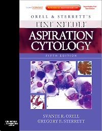
Orell and Sterrett's Fine Needle Aspiration Cytology, 5th Edition
Expert Consult: Online and PrintOrell & Sterrett’s Fine Needle Aspiration Cytology 5e provides you with a logical and systematic approach to the acquisition, interpretation and diagnosis of FNA biopsy samples. It is an ideal resource for all those requiring an authoritative and systematic review of the cytological findings in those malignant and benign lesions likely to be the target of FNA. The book is lavishly illustrated with high quality colour images that demonstrate the cytological features as well as their relevant immunohistochemical and molecular findings. Organized into anatomical regions, each chapter is consistently organized into two parts: the first deals with clinical and technical aspects followed by a systematic presentation of cytological findings. This is your perfect practical bench resource for daily reference in the laboratory.
Orell & Sterrett’s Fine Needle Aspiration Cytology 5e provides you with a logical and systematic approach to the acquisition, interpretation and diagnosis of FNA biopsy samples. It is an ideal resource for all those requiring an authoritative and systematic review of the cytological findings in those malignant and benign lesions likely to be the target of FNA. The book is lavishly illustrated with high quality colour images that demonstrate the cytological features as well as their relevant immunohistochemical and molecular findings. Organized into anatomical regions, each chapter is consistently organized into two parts: the first deals with clinical and technical aspects followed by a systematic presentation of cytological findings. This is your perfect practical bench resource for daily reference in the laboratory.
New to this edition
Brand new chapter on cytological findings in infectious diseases.
Inclusion of immuno-profiles and other relevant ancillary tests.
New illustrations.
New contributing authors.
Available online via Expert Consult.
Key Features
Provides practical tips and advice on how to avoid pitfalls and ensure accurate diagnoses.
Over 1,200 colour illustrations capture each entity’s cellular, morphological and immunohistochemical appearance.
Chapters have been up-dated and revised and a brand new one on cytological findings in infectious diseases added.
Both MGG and Pap smears illustrated in parallel as well as the corresponding histology to help provide side-by-side analysis.
Access the full text online and download images via Expert Consult.
Author Information


