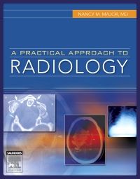
This lavishly illustrated book is a perfect introduction to the exciting field of radiology. A lively, easy-to-read writing style explores the diagnostic imaging issues associated with each body region. Extensive tables, color-coded key points, and detailed drawings make finding essential information a snap. Case studies and high- resolution images demonstrate radiologic principles and characteristic findings for a wide range of disease entities. All imaging modalities are covered including fluoroscopy, CT, MR, nuclear medicine, and more. An ideal text for anyone interested in diagnostic imaging, A Practical Approach to Radiology not only gives you the information you need, it brings it to life.
Key Features
- Features a full-color, templated format, making pertinent information easy to digest and recall.
- Includes color-coded tables that discuss differential diagnoses and indications for types of exams.
- Uses key points to list the findings on an x-ray as well as outlining search patterns.
- Explores the latest, cutting-edge technologies in the field, including spiral CT and 3-D reconstruction.
- Contains over 800 images to demonstrate important points.
Author Information
By Nancy M. Major, MD, Professor of Radiology and Orthopedics, University of Colorado School of Medicine, Director of Imaging, Sports and Performance Center, Boulder, Colorado
1. Basic Concepts of Imaging Methods
2. Chest
3. Abdominal Imaging
4. Musculoskeletal Imaging
5. Neuroimaging
6. Pediatric Imaging
7. Interventional Radiology
8. Nuclear Imaging
9. Breast Imaging


