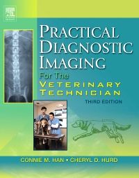
A complete and practical guide, this text describes how to produce high-quality radiographic and ultrasound images. The first half of the book covers equipment, safety, and technique - all major responsibilities of the veterinary technician. The second half details radiographic positioning for small animals, large animals, and exotics. Reflecting the major role of ultrasonography in veterinary practice, the book concludes with an expanded chapter on diagnostic ultrasound.
New to this edition
New cardiac ultrasound scanning techniques with 20 new ultrasound images
New images of the latest x-ray equipment
Updated pedagogical features, including outlines, key points, chapter objectives, and helpful hints for veterinary technicians
Key Features
- Practical, concise clinical format
- Written at the appropriate reading level for technicians
- Abundant illustrations emphasize basic radiographic and ultrasonographic principles, techniques, and equipment used in veterinary practice
- Concise and understandable discussion of physics and radiography
- Examples of common artifacts show how to avoid misinterpretation of imaging studies
- Excellent coverage of radiation safety
- Practical technique charts
- Excellent coverage of small- and large-animal positioning
- Exotics chapter featuring rodents (including ferrets), reptiles, and birds
- Ultrasound chapter that includes normal ultrasonographic findings, along with corresponding images
- Helpful hints given for obtaining quality images and avoiding common pitfalls reflect the authors' experiences at a busy teaching institution
Author Information
By Connie M. Han, RVT, Veterinary Teaching Hospital, Purdue University, West Lafayette, IN and Cheryl D. Hurd, RVT, Veterinary Teaching Hospital, Purdue University, West Lafayette, IN
PART ONE: RADIOGRAPHIC TECHNIQUE AND EQUIPMENT
1. X-ray Generation
2. Achieving Radiographic Quality
3. Exposure Variables
4. Recording the Image
5. X-ray Equipment
6. Darkroom Techniques
7. Radiation Safety
8. Developing a Small Animal Radiographic Technique Chart
9. Radiographic Artifacts
PART TWO: RADIOGRAPHIC POSITIONING
10. Small Animal Radiography
11. Basic Small Animal Dental Radiography
12. Contrast Studies
13. Exotic Animal Radiography
14. Large Animal Radiography
15. Diagnostic Ultrasound


